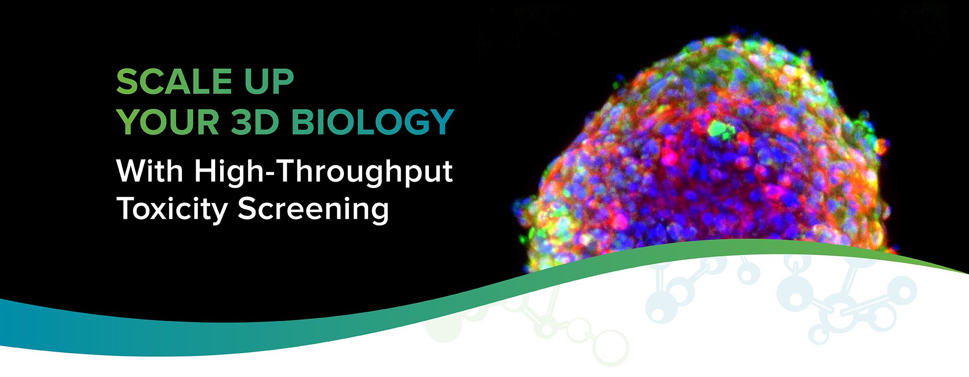
Explore our fully-integrated, automated 3D biology workflow solution for toxicity evaluation
Measuring compound effects on human cardiac tissues is a time-consuming and resource-heavy process. If you are looking for a 3D toxicity screening solution, we can help you build a personalized, end-to-end workflow to deliver robust, reproducible results at scale. Using 3D cell models enables your assays to have greater predictivity and ultimately lower the failure rates later in the drug development process.
The following workflow can be used during drug discovery and development to screen compounds for potential cardiotoxic or neurotoxic effects. While the FLIPR Penta allows for exceptionally fast, high-throughput measurements of calcium oscillation patterns, the ImageXpress High Content Imaging System allows for the fast acquisition and analysis of complex biological samples.

Do you want a complete workflow for toxicity screening?
Learn more about the cardiotoxicity screening workflow
Structural organization and functional analysis of compound responses in 3D human iPSC-derived cardiac tri-culture microtissues
In this video, Simon Lydford, Senior Application Scientist at Molecular Devices, presents his poster on the aforementioned workflow, explains why 3D cell models are becoming more common and how the FLIPR Penta system can perform functional assays on such models, such as cardiac microtissues.
Structural organization and functional analysis of compound response in 3D human iPSC-derived cardiac tri-culture microtissues
In this application note, we highlight the utility and biological relevance of using iPSC-derived cell types in 3D micro-tissues as a promising model for measuring compound effects on human cardiac tissues in a high-throughput format.
Structural organization in 3D human iPSC-derived cardiac tri-culture microtissues
In this study, we used a tri-culture model created by mixing iPSC-derived cardiac cells with primary adult fibroblasts and iPSC-derived endothelial cells. A variety of parametric responses were demonstrated using a set of 8 known modulators of cardiac activities. The assay can be used to test developing drugs and screen chemicals for potential cardiotoxic hazard.


